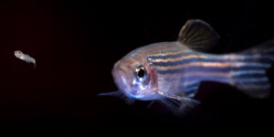


Cut a zebrafish’s heart and something remarkable happens. Within seconds, the fish clots the wound and stops the bleeding. Cells start to divide to make new heart muscle and blood vessels. Two months later, the zebrafish has completely regenerated its heart. If only we could do that.
It turns out that zebrafish’s heart resembles that of the human heart so closely –70% of human genes are also found in the fish – it has made it a relevant and compelling model for research into how the heart develops and breaks.
Part of the draw is that zebrafish are transparent early in life. But by adulthood, the fish are mostly opaque, rendering the live observation of heart regeneration nearly impossible. But thanks to a novel device built by Professor Megan McCain and her Ph.D. student Joycelyn Yip at the USC Viterbi School of Engineering’s Laboratory for Living Systems Engineering, scientists are now able to keep hearts alive and functional outside of the fish longer than ever before.
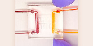
McCain and Yip designed and manufactured a square device, slightly larger than a quarter, for maintaining explanted adult zebrafish hearts that continuously supplies the hearts with nutritious fluid to keep them alive. The device is also compatible with live imaging. Crucial to their success was a partnership with Dr. Ching-Ling Lien and Dr. Michael Harrison, experts in zebrafish heart physiology at the Heart Institute and the Saban Research Institute, both at Children’s Hospital Los Angeles (CHLA). Their collaboration was also supported by The Saban Research Institute intramural funding (Team Science Award) that encourages convergence and team building. They jointly published a paper in the journal Lab on a Chip.
“Conventionally, our collaborators in the Lien lab would remove the heart from the fish, injure it, put it into a dish, and image whatever regeneration can be observed,” said McCain, a Chonette Early Career Chair and assistant professor of biomedical engineering, stem cell biology and regenerative medicine. “But the heart would not survive more than three to five days, which is not long enough for complete regeneration.”
With their device, the team was able to keep the fish hearts beating for one to two weeks. Using time-lapse imaging, they were also able to visualize coronary vessels developing from the inner lining of an injured heart, providing new blood supply to the site of injury.
“This collaboration with Megan McCain’s lab using the engineered device helps us push the imaging capacity of regenerative model organisms to a new level,” said Dr. Ching-Ling Lien, associate professor of surgery at the Keck School of Medicine of USC.
Made mostly out of PDMS, a silicon-based organic polymer, the device was embedded with a fluid path comprising inlet and outlet reservoirs, a network of channels, and four triangular compartments that are slightly larger than an average adult zebrafish heart.
“Another challenge with studying the hearts in a dish is trying to image the same spot over a long period of time, because the hearts are very small and move around in the dish,” said Yip whose background in mechanical and biomedical engineering informed the prototyping of the device. “We designed triangular chambers that nestled the hearts into a fixed place so that they can be moved on and off a microscope stage while keeping their orientation intact.”
McCain and Yip are now focusing their efforts on refining their device to extend the time they can keep zebrafish hearts alive, which could reveal even more about how this heroic aquarium fish renews its heart from the inside out.
“My lab mostly makes devices to culture heart cells. This project was about applying those techniques and tools to culture an entire organ,” said McCain, whose work with engineering organs-on-chips with human heart cells is paving the way for a dramatic testing ground for new drugs, as well as a miniature lab for personalized pharmaceutical testing.
Heart disease is the No. 1 killer in the U.S. and in the world, having claimed more lives in 2019 than the first and second world wars combined.
“Establishing these methods and tools is the first step towards potentially translating mechanisms of zebrafish heart regeneration to human heart repair, which would be a completely new treatment strategy” McCain said.
Published on February 14th, 2020
Last updated on February 14th, 2020