

Algae. Photo/ Anni Roenkae.
Tiny hairlike organelles called cilia cover many cells from single-celled microorganisms to mammalian tissue. Motile cilia beat actively generating cilia-driven flows.
In aquatic species, cilia commonly occur along external and internal surfaces, where they have a broad array of functions, from food capture to acquisition of microbial partners. When animals invaded terrestrial habitats, cilia became more internalized along the outer surfaces of animal tissue —in the lungs, in the brain, in the reproductive tract — rendering them difficult subjects for direct study. In a healthy body, motile cilia coordinate with each other to achieve specific modes of motion, so they can direct flow in a certain direction. Practically, this might appear as mucus being pushed from the lungs or sperm being helped along the reproductive tract to meet with an egg.
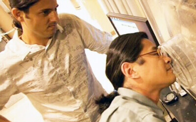
It is these flows—and the shifts between them—that are essential to cilia’s role as the first line of defense in the body. Eva Kanso, Zohrab A. Kaprielian Fellow in Engineering and professor and post-doctoral scholar Yi Man, both in the Department of Aerospace and Mechanical Engineering, performed computational experiments to better understand how transitions between different modes of ciliary coordination occur.
The researchers asserted that these transitions occur in one of two ways: changing the level of coupling between cilia or shifting the activity level of the cilia. Results were published in Physical Letters Review and highlighted as an Editors’ Suggestion.
Kanso said that cilia, in mammalian tissue, are in important places—like the upper respiratory tracts, where they form the first line of defense against environmental particles, viruses and bacteria—making understanding how they work that much more important.
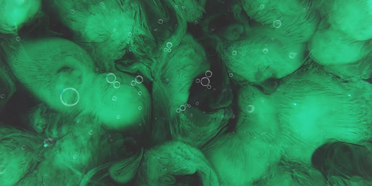
However, owing to the difficulty of accessing cilia in these locations, researchers often use aquatic microorganisms as model system to get insight into cilia motion. This is possible because cilia structure is conserved across species. Knowledge gained from studying the motion of cilia in algal cells, for example, could translate to direct insight into cilia behavior in the human body.
“If the cilia are not coordinated, each is moving in a different direction. Their purpose is to push fluids and if every cilium is working against every other cilium, they won’t be able to achieve this in a consistent, coherent manner,” Kanso said.
But beyond coordination, Kanso said, the next biggest consideration is how the cilia transition from one coordinated mode of motion to the next. “The fate of the cell is dependent on these transitions.”
How Transitions in Cilia Coordination Provide Vital Diagnostic Information
The study Kanso and Man conducted focused on the interaction of a pair of cilia. In the future, they hope to look at a whole ciliary bed, with the hopes of further understanding not only how cilia function, but also the greater implications for how their coordination reflects on the health of an organism.
Kanso said that the distance between cilia—one factor her study suggests change transitional behavior in cilia—can be impacted by certain diseases, for example lung disease. Given this, her team is hoping to translate the tools and techniques they acquire from a broader study of ciliary beds to work with Dr. Amy Ryan, assistant professor, and Dr. Janna Nawroth, senior research scientist, both at the Keck School of Medicine at USC.
“The long-term vision is that maybe there will be some way to not just look at individual cilia but grow ciliated tissue—for instance based on a swab of human cells—and to look at its coordination. How well the cells coordinate could be used as a diagnostic tool to better understand how far a disease has progressed and what a patient’s prognosis is,” Kanso said.
What Happens When Cilia Move
Cilia are made of a system of microtubules. There are molecular motors that “walk” on those microscopic tubule structures—or, in other words, “they bind and unbind,” Kanso said. The movement of cilia are largely caused by these motors, she said, as they generate forces, which in turn produce oscillatory motion.
As stated above, the researchers found one of the main triggers for transitions in these coordinated movements to be changes in activity level among the cilia.
“Activity of the cilia is dependent on certain molecules in their environment, for example calcium can impact this. In return, this can change the coordination mode in the cilia,” Kanso said.
For example, in the lungs, a shift in the environment—perhaps the result of diseased tissue—could trigger a shift in the activity level of the cilia. As a result, the cilia may be less coordinated and this transition to an uncoordinated state could be detrimental for the health of the lung. Without appropriate motion sweeping harmful bacteria and particles out of the lung, it may build up within the lung. In turn, this could change the cilia’s environment, causing a feedback loop that continues to suppress normal cilia function, Kanso said.
This work was funded by the National Science Foundation.
Published on October 20th, 2020
Last updated on October 21st, 2020