

Illustration: Dall-E
At the Alfred E. Mann Department of Biomedical Engineering‘s 50th anniversary symposium Sept. 3, Dr. Charles Liu delivered the keynote address for the neuroengineering session with a provocation that landed differently coming from a neurosurgeon: the reason the most powerful brain technologies rarely reach patients is not a science problem. It is a proximity and access problem.
Liu is a professor of clinical neurological surgery, surgery, psychiatry and behavioral sciences, and biomedical engineering at the Keck School of Medicine of USC and USC’s Viterbi School of Engineering, director of the USC Neurorestoration Center, and director of the USC Comprehensive Epilepsy Care Consortium. He trained first as a chemical engineer, earned a PhD at Rice University, then went to Yale for his MD.
“I went to medical school so I could be a better engineer,” he said.
When Engineers Are in the Room
The concept Liu has spent years building is what he calls a living laboratory: not a call for more inefficient collaboration between hospitals and universities, but a structure in which engineers are welcomed to work physically inside clinical environments — in the operating room, in the ICU — so the problems they solve are the ones patients actually have.
The USC Neurorestoration Center, where Liu serves as founding director, is the embodiment of that idea. The clinical venues include Rancho Los Amigos National Rehabilitation Center in Downey, California, where Liu serves as Chief of Innovation and Research, Casa Colina Hospitals, where Liu serves as Medical Director of the Research Institute, as well as Keck Medical Center of USC, and other hospitals as far away as the Central Valley.
Working alongside clinicians there, USC engineers, as well as collaborators from Caltech and UC Irvine, have unprecedented access to human patients and problems they need solved. For example, Liu and Vasileios Christopoulos (Assistant Professor of BME and Neurological Surgery) have conducted the first applications of functional ultrasound imaging of the human spinal cord, as well as the brain of patients undergoing epilepsy surgeries.
With collaborators from Caltech, functional brain imaging using photoacoustics and functional ultrasound have been successfully achieved in patients whose skulls have been surgically removed and in some cases subsequently reconstructed using “transparent” implants — a population that offers a rare window into brain function that intact skull ordinarily blocks.
Those patients are now the proving ground for something more ambitious.
The Bone Is the Problem
Medical imaging, transformative for patient care, began with X-rays. Virtually every part of the human anatomy has been imaged with X-rays except for the brain because the skull makes the organ effectively opaque. The same applies to the spinal cord, which has even more limited options because even CT and MRI give worse image resolution than even that of the brain. Many other low-cost imaging tools that work everywhere else in the body also have limited application in the nervous system because of the bone that encases it for protection.
Patients who have undergone or is undergoing neurosurgery to remove part of the skull or spinal lamina— for tumors, trauma, or swelling — briefly lose that barrier, and Liu’s group, working with engineers, has maximized this unique opportunity to use these windows to validate imaging techniques that could eventually work without removing bone at all. In fact, Liu is actively working with Mack Yaoheng Yang (Assistant Professor of BME) on ways to render intact human skulls “transparent” to ultrasound.
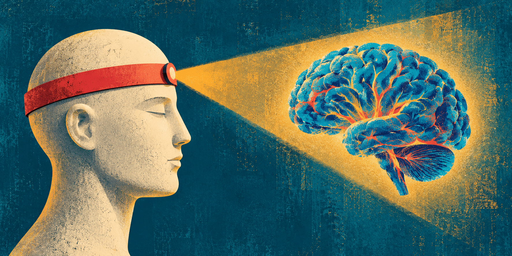
The most concrete result is a wearable photonic device, developed with Caltech collaborators, designed to measure brain blood flow and assess stroke risk without a conventional scanner.
The approach is deliberately simple: a patient holds their breath, a brief stress on the brain’s circulation that is analogous to the treadmill test a cardiologist uses to reveal hidden heart disease. A headband worn during that breath-hold measures how blood flow responds. Liu and collaborators have found that response correlates with stroke risk and can detect brain injury in environments far beyond traditional hospitals.
The explicit goal is scalability. Liu described wanting the components to cost low hundreds of dollars, sourced from consumer electronics suppliers — a direct contrast with the six to seven-figure scanners that currently anchor brain monitoring in well-resourced hospitals. Testing started in neurosurgery patients but has already been expanded to patients with intact skulls.
“The best part is that the patients actively participate in finding solutions to their most challenging problems working with the most capable engineers with superpowers,” Liu said.
Translation Requires Presence
The headband is one output of a larger argument Liu has been making throughout his career: that clinical translation stalls not because the scientific principles do not apply, but because the engineers are too far from the patient. The living laboratory is his answer — a model in which the distance between where the research happens and where the patient is being treated has been reduced, in some cases, to the same room including the operating room.
“The solutions to medicine’s biggest problems,” he said, “will be found by allowing the engineers wielding superpowers to come as close as possible.”
Published on September 16th, 2026
Last updated on September 16th, 2026