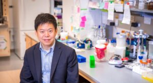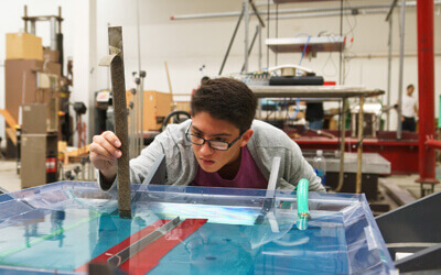


Cristina Zavaleta, an assistant professor in biomedical engineering, will join the USC Michelson Center for Convergent Bioscience when it opens this November. Photo credit/Valentina Suarez
Cristina Zavaleta, a self-proclaimed Disney fanatic, is a firm believer in the gumption expressed by one very famous animator:
“If you can dream it, then you can do it.” – Walt Disney
Zavaleta, a Gabilan Assistant Professor of Biomedical Engineering, is a big dreamer and do-er in her own right.
Her bailiwick: nano-based molecular imaging, a tool that can be exploited in early cancer detection and tumor resection, or surgical removal.
Her expertise makes for a superb fit as a faculty member in the USC Michelson Center for Convergent Bioscience, a 190,000 square-foot, high-tech research facility supported by a $50 million gift from retired spinal surgeon Dr. Gary K. Michelson and his wife, Alya Michelson. By drawing from a diverse network of scientists, engineers and students, the new center fosters biomedical discovery, innovation and real-world solutions to fast-track detection and cures for diseases ranging from microbial infections to Alzheimer’s and cancer.
Nanoparticle Know-How
Zavaleta’s post-doctoral fellowship in the Molecular Imaging Program at Stanford University exposed her to a plethora of imaging tools. She plans to bring her specialized knowledge to the Michelson Center and hopes to help answer some important questions related to earlier cancer detection. Her research is currently focused on developing new nano-based imaging contrast agents.
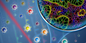
Zavaleta explains that nanoparticles are ideal to ferret out the earliest signs of cancer. They can be engineered to glow brighter and circulate longer in the bloodstream than small molecule contrast agents. Another plus, their size – roughly one hundred times smaller than our own cells – which make them more likely to passively accumulate and be retained within tumor areas. The result: an image that glows in tumor areas where the enhanced nanoparticles are present.
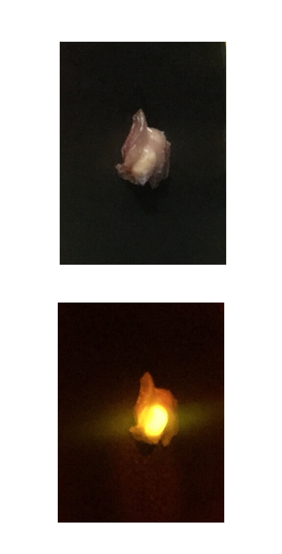
Top: Excised tissue from a mouse after intravenous injection of glowing nanoparticles. Bottom: By using the appropriate light and filters, the tumor area glows, showing precisely where the tumor is surrounded by normal non-glowing tissue. Photo courtesy/Cristina Zavaleta
Better still, the nanoparticles can be modified to actively target a specific tumor area.
“You can decorate the outside of the nanoparticles with different proteins or antibodies to make them actively target something specific, like certain biomarkers overexpressed on the tumor cell’s surface.” Zavaleta said. “By doing this, we’re actually using the biology and mechanics that our own cells already use.”
A Paint for Tumors?
In addition, the nano-based imaging can be used by surgeons to “paint” a tumor’s footprint, which would be visible on a screen in the operating room to help ensure all tumor margins are cleared during surgical removal.
“If a tumor margin is missed during surgery, it can be detrimental to the patient, possibly leading to recurrence and repeat surgery,” Zavaleta said. “Unfortunately, pathology takes several days, and we only find out later if some of the tumor was missed after surgery.”
An Imaging Ace
An expert in several biomedical imaging tools, Zavaleta’s arsenal includes ultrasound, CT, MRI, PET, SPECT, fluorescence, photoacoustics, and Raman imaging devices.
“Each of us at the Michelson Center offers a different set of tools to answer some important scientific questions,” Zavaleta said. “I’m excited to bring what I call my ‘imaging tool box’.”
Each of us at the Michelson Center offers a different set of tools to answer some important scientific questions. I’m excited to bring what I call my “imaging tool box.”Assistant Professor Cristina Zavaleta
Each imaging tool has its own advantages and disadvantages in cancer detection. While some are more sensitive, others have better spatial resolution. Some devices even have multiplexing capabilities, or the ability to image multiple molecular processes simultaneously. And depending on the type of image required, Zavaleta knows which tool to employ.
“CT can offer you great structural information about the attenuation of the x-rays through the body, so you will see broken bones or masses within the body with high spatial resolution,” Zavaleta said. “MRI offers great soft tissue contrast as well, particularly in the brain, and ultrasound is a safe, quick and inexpensive imaging technique that can offer important high resolution structural images along with real time blood flow information.”
What’s Your Function?
While most of these devices are used to look at structural details, Zavaleta says functional information is gleaned through the addition of the glowing molecular contrast agents.

Still microscopic image of a real time video showing glowing nanoparticles (green) traveling though the blood stream (red). Photo courtesy/Cristina Zavaleta
“The contrast agents we are engineering in the lab are going to be nanoparticles that have something on their surface that interacts with the body on a molecular level, giving us important functional information,” Zavaleta said. “One of the most commonly used functional imaging contrast agents is FDG or fludeoxyglucose.”
FDG is a glucose analog that gets processed in the body like regular glucose during metabolism. Areas of the body that have cancer will have high metabolic activity, taking up more and more FDG and creating brighter imaging in those regions.
Lighting a Path Forward
Zavaleta looks forward to tapping the myriad resources at the Michelson Center – not only human talent, but also cutting-edge technology.
“I’m very excited about working with Scott Fraser’s group,” Zavaleta said. “Reason being, they have some of the most awesome optical imaging tools that exist on the planet, and several of them can answer very important questions about our contrast agents.” Fraser is the Elizabeth Garrett Chair in Convergent Bioscience.
She is also eager to utilize the CEMMA facility, to be newly housed in the Michelson Center, which will feature several state-of-the-art electron microscopes.
Equally attractive to her is the opportunity to be a part of something from the very beginning, or “getting in on the ground floor,” she says.
Armed with her high-tech toolbox, Zavaleta is poised to play a vital and convergent role at the new Michelson Center, in a way that brings to mind a different quote by the beloved animator previously quoted. And it just so happens to pay due to her unique skillset:
“Of all our inventions for mass communication, pictures still speak the most universally understood language.” – Walt Disney
About Cristina Zavaleta
Joining USC in Fall 2017, Zavaleta is the newest addition to the core faculty of the Department of Biomedical Engineering. The Southern Texas native completed her undergraduate work in nuclear medicine at the University of the Incarnate Word in San Antonio. She went on to receive her Ph.D. in medical physics from the University of Texas Health Science Center, also in San Antonio, where she was introduced to biomedical nanotechnology research.
Published on October 31st, 2017
Last updated on April 7th, 2022