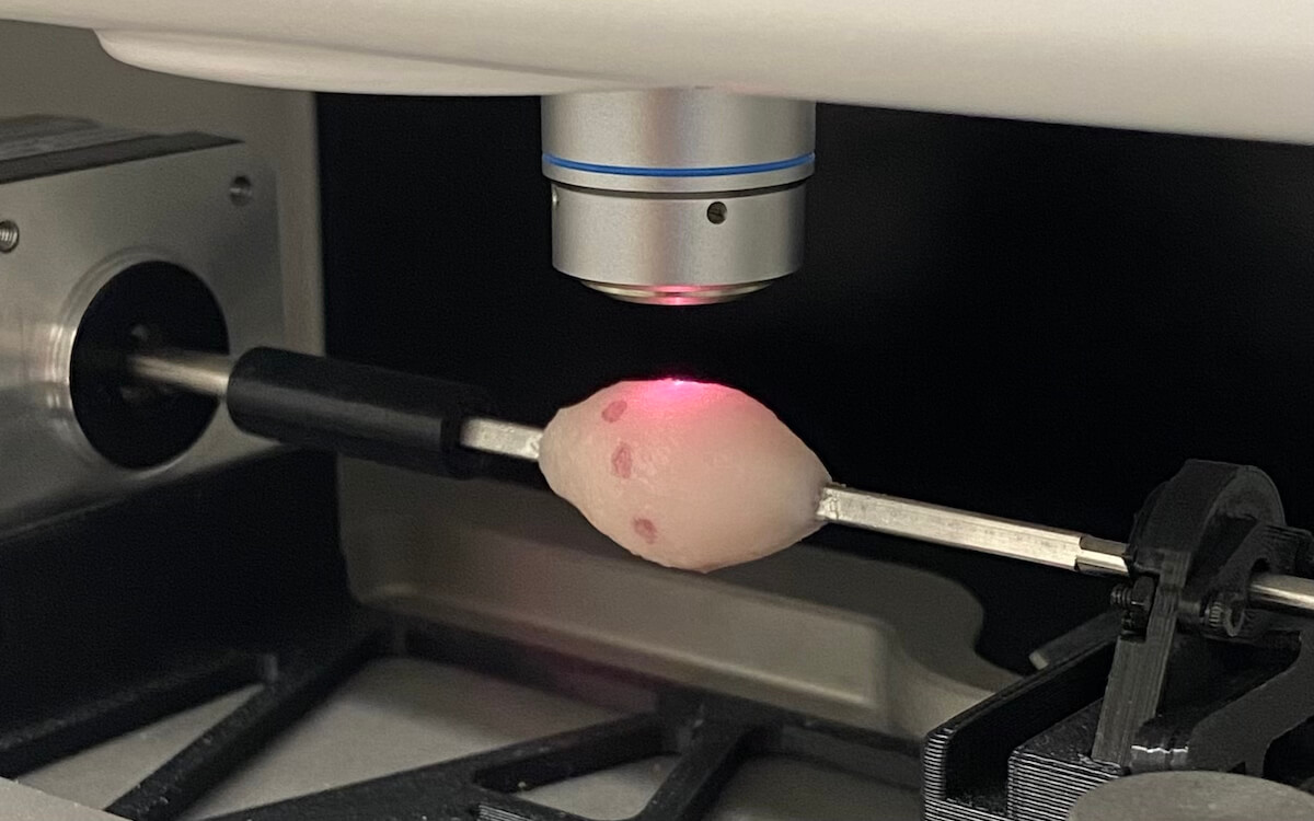
The Zavaleta Lab’s Raman Rotisserie device creates a map of the surface of a resected tumor to aid surgeons in the operating room. Image/Alex Czaja
Like many Texans, Cristina Zavaleta grew up enjoying the culinary delights of the state’s famous smokehouse BBQs. She couldn’t have imagined that those humble rotisseries of her childhood would one day inspire a game-changing device for the operating room that could help surgeons prevent tumor recurrence.
On a team excursion to Disneyland, the WiSE Gabilan Assistant Professor of Biomedical Engineering and her students were reminded of rotisseries when they encountered a food vendor at the Star Wars-themed land, Galaxy’s Edge. It was a lightbulb moment. The rotisserie configuration was a perfect way of intricately scanning excised tumors, with the help of the Zaveleta Lab’s unique nanoparticles, to light up where the cancerous tissue may not have been entirely removed from the patient. Surgeons could then be guided to precisely remove the remaining tumor, all while the patient is still under anesthesia. The result would reduce the need for traumatic repeat surgeries and potential cancer recurrence and metastasis.
Zavaleta and her team built the device, which they dubbed the Raman Rotisserie. It physically rotates a tumor specimen and works in conjunction with an imaging technique known as Raman spectroscopy, which scans the surface of the excised tumor. Their research, which aims to improve the success rate of breast cancer lumpectomies, has now been published in NPJ Imaging.
“We describe it to people as being a rotisserie, because I think it helps them understand that it’s on a spit, and it’s rotating. And the rotation allows us to survey the entire surface of the sample we’re looking at,” Zavaleta said.
“If we start at the very beginning – The problem is that surgeons need additional help ensuring that they’ve actually taken out the entire tumor. So, in that sense, having something that can interrogate the surface of the resected specimen would be helpful,” she said.
Zavaleta said that to achieve a successful resection, the surgeon must cut the cancerous tissue while keeping it encapsulated in an unbroken layer of regular healthy tissue. If that encapsulation is incomplete or broken, it could mean that parts of the tumor remain inside the patient.
The current best practice for surgeons to ensure complete tumor removal, particularly when it comes to lumpectomies in breast cancer patients, is to locate the tumor via mammography and then remove it. However, the surgeon primarily relies on visual cues and tactile feedback to cut the tissue. The surgeon must then send the resected sample away to a pathology lab for imaging to ensure it has been removed completely with “negative tumor margins.” As such, patients must wait numerous anxious days or up to a week to see if their surgery was successful. If not, they could face follow-up repeat surgery. The Zavaleta Lab’s device is designed to complete this process in under an hour.
“The possibility of repeat surgery is devastating, so having a tool like this could potentially help surgeons. It could all be done right there, adjacent to the operating room. Before sending the tissue to pathology, the surgical team could just place the prepped tissue in the device, spin it, image it, and then make the decision right there in-house,” Zavaleta said.
Because the rotisserie device helps support surgical accuracy, it also enables improved conservation of healthy tissue, meaning breast cancer patients could more confidently opt for lumpectomies rather than total mastectomies.
The imaging process works in conjunction with the Zavaleta Lab’s gold-core nanoparticles that are patterned with 26 different biomarker targets — highlighting the behavior of cancer tissue at a molecular level.
The surface of the resected tumor tissue would be coated with the nanoparticles before being placed on the rotisserie and imaged using Raman spectroscopy, which creates a 3D map of the surface of the tumor. Any break in the margin of the healthy tissue that leaves the cancerous tissue exposed would then be “lit up” by the nanoparticles, which work like a coordinate system that alerts the surgeon to which parts of the tumor may remain in the patient.
“Our strategy is unique in that it uses a multiplexed panel of nanoparticles capable of targeting various tumor biomarkers simultaneously to improve our ability to specifically identify and localize the residual tumor margins. We’re also utilizing the principles of Raman scattering to generate our images. What that affords us is the ability to interrogate multiple cancer biomarkers at the same time by using our different ‘flavors’ of targeted nanoparticles where we can assign different colors to localize these distinct targets,” Zavaleta said.
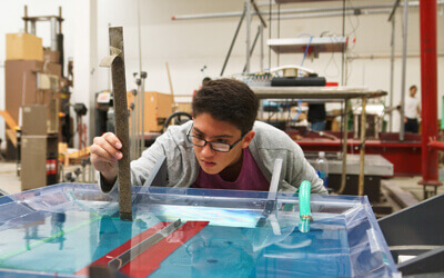
When it came time for the team to create the prototype, graduate student in the Zavaleta Lab, Alex Czaja, took a visit to Williams Sonoma to study the design of kebab skewers. The team needed an alternative to the cylindrical shape to ensure the tumor sample didn’t slip during rotation. It turned out that the sharp edges of kebab skewers created a solid base to mount tissue on so that it remained fixed in place on the rotisserie accessory device.
“This process was fun because it was an opportunity for both my graduate and undergraduate students to come up with very creative ways to execute this,” Zavaleta said. “Our undergrads, Alice Jiang and Matt Blanco, were an integral part of the problem-solving process.”
The research team is now working on improving the speed of the rotisserie to sample a tumor’s surface area with greater efficiency. They are also looking to work with clinical collaborators to begin testing the prototype’s effectiveness using human tissue samples.
“There has been a lot of excitement in the field for image-guided tumor resection in general. We’ve spoken to surgeons who are very excited about any sort of tool to help give them some assurance that they’ve been successful with resecting the entire tumor,” Zavaleta said. “With our tool, we’re offering a quick imaging solution while the patient is still in the operating room that can give physicians actionable clinical feedback.”
Published on April 4th, 2024
Last updated on July 23rd, 2024
