

New MRI machine being moved into USC Michelson earlier this year/Photo Credit: Ben Paul
Researchers at USC’s Dynamic Imaging Science Center (DISC) have been able to produce a clear scan of fetal cardiac anomalies for expectant mothers who had previously not been able to see their fetuses clearly. The technique, which relies on a customized algorithm and customized low-field MRI machine, would be an important diagnostic tool for the estimated one percent of babies born with congenital heart cardiac defects. Researchers who presented the paper at the “Annual Scientific Sessions of the Society for Cardiovascular Magnetic Resonance,” explain that “fetal cardiac MRI can be useful when ultrasound imaging is suboptimal or limited.” The researchers from USC along with co-authors from Children’s Hospital of Los Angeles (CHLA) believe this new method can provide enhanced imaging and clearer pictures of fetal heart disease for patients who were not able to get a clear picture through traditional ultrasound imaging.
The new method for fetal MRI imaging can be applied safely, say the researchers. While the algorithm that created these methods is custom, the researchers want to make it available to help people all over the world as they believe such a tool may be able to help doctors proactively address the congenital anomalies that “account for 20 percent of infant deaths.” DISC, directed by scholars from the USC Viterbi School of Engineering, and the low-field MRI machine customized by Siemens Healthineers with input from USC researchers, has been in place at the USC Michelson Center for Convergent Bioscience since 2021. This innovative technology improves the imaging of moving organs, such as the heart, lungs, and joints, to make diagnoses and treatment more accessible.
“This MRI,” says the paper’s lead author and post-doctoral scholar at USC Viterbi DISC, Ye Tian, “…can provide more data with less risk.”
“The low field magnet excels at real-time (moving) imaging, which is critical because fetuses are constantly moving in utero.” said co-author John Wood, who is the Director of Cardiovascular MRI at the Children’s Hospital Los Angeles, and Professor of Pediatrics and Radiology at USC.
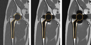
This particular MRI at USC is one of only three such machines in world. It includes all the technologies of a traditional MRI scanner while utilizing the advantages of a field strength that is three to six times lower than conventional systems, for unique imaging capabilities. With this machine, DISC explores real-time imaging of traditionally difficult areas, e. g. in speech production or sleep apnea therapy and for patients who had oral cancer resection. Additionally, the new low-field MRI, is allowing researchers to study orthopedic metal implants and how these implants affect surrounding tissue.
For USC, DISC and its research builds on decades of imaging at the USC Viterbi School of Engineering—including at the school’s Signal Image Processing Institute, where early work became the basis of the JPEG and MPEG standards for still and video image compression and transmission.
Published on March 29th, 2023
Last updated on April 4th, 2023