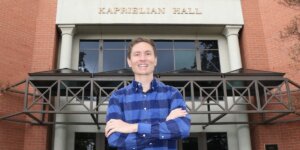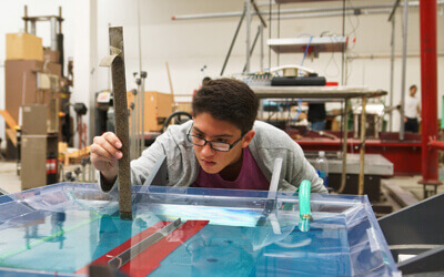


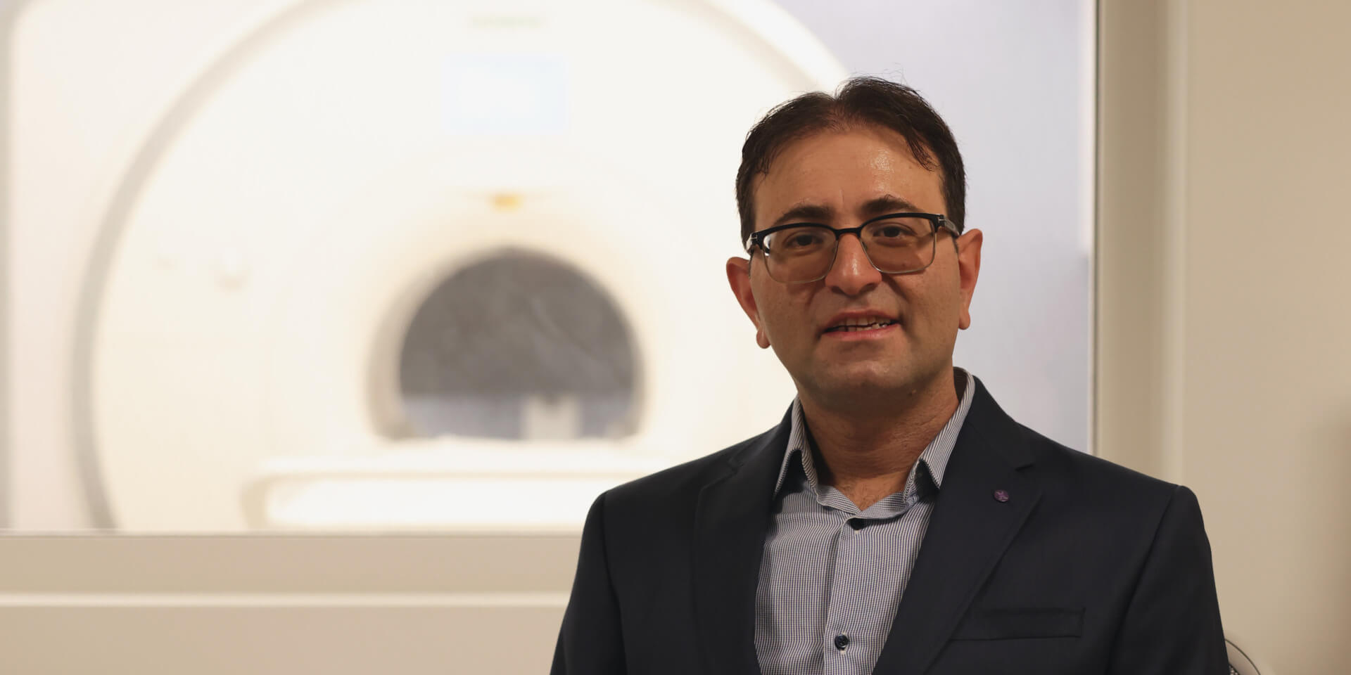
Mahdi Soltanolkotabi with a low-field MR machine in the USC Dynamic Imaging Science Center. (USC Photo/Xenia Xu)
What if you wanted to create vivid, professional-quality photos, but all you had at your disposal was your old, four-generations-ago smartphone? That’s basically the kind of imaging problems Mahdi Soltanolkotabi is working on. Except the pictures he reconstructs could alleviate suffering for patients with uncommon nerve disorders.
Soltanolkotabi, an associate professor of electrical and computer engineering, computer science, and industrial and systems engineering, is among the recipients of the National Institutes of Health Director’s New Innovator Awards announced Tuesday. The $2.4 million research grant is part of the NIH’s High-Risk, High-Reward Research program “to support highly innovative scientists who propose visionary and broadly impactful behavioral and biomedical research projects.” The last ECE recipient of the prestigious award was Maryam Shanechi in 2020.
Soltanolkotabi’s project develops new algorithms powered by artificial intelligence to augment scans produced by magnetic resonance imaging (MRI) machines to improve their resolution. The goal is to take scans made by “low-field” machines and build them into higher-quality images so they can be of the most use to radiologists interpreting the exams.
Scanners of increasing power and sophistication have been used at medical centers in recent years. Their magnetic field strength is measured in units called Teslas: A prized 7-Tesla Siemens was installed at the USC Stevens Neuroimaging and Informatics Institute in 2017, and the FDA approved it for clinical use the next year. But not every patient has access to one. Much more plentiful are less-powerful devices down to the 3T and 1.5T level. A patient who has a pacemaker, or a surgical implant with a metal component, is limited to using a low-field scanner such as the one recently installed in the USC Dynamic Imaging Science Center (DISC). These implants are either unsafe at higher-field strength magnets or cause significant artifacts, or anomalies, that hinder image interpretation.
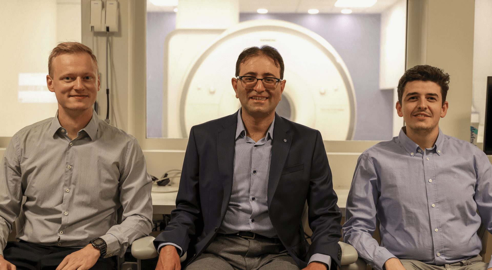
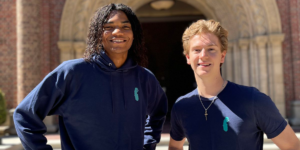
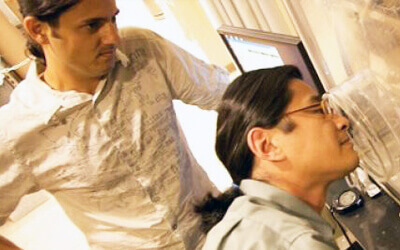
Dr. Soltanolkotabi (center) with Zalan Fabian (left) and Berk Tinaz. (USC Photo/Xenia Xu)
The goal of Soltanolkotabi’s work is to reconstruct images from a low-field machine so that they’re every bit as revealing as the images from a more powerful machine. The result is reduced cost – low-field scanners can potentially be so small they’re portable, rolled in on castors – as well as improved image quality.
“The low-field scanner gives you low-quality images,” Soltanolkotabi said. “The question is, can we take low-field measurements but algorithmically compensate for them, as if you had high-field measurements? The dream scenario is to have a portable MR device but get as high quality of a reconstruction as if you had a bulky high-field scanner.”
The nerve disorder Soltanolkotabi is working on is called brachial plexopathy, which happens when there has been damage to the brachial plexus, the bundle of nerves that run from the lower neck through the upper shoulder area. Such an injury can cause severe shoulder and upper arm pain or weakness. Nerves can be hard to see in great detail, so the kind of heightened level of clarity one would get with a high-field scanner helps radiologists make more informed decisions on the specific type and location of injury. This in turn informs the surgeon on management strategies.
“It tells the surgeons whether they should operate or not,” says Maryam Soltanolkotabi, a musculoskeletal radiologist at the University of Utah and a collaborator of Mahdi Soltanolkotabi’s. “This is one example where the radiologist would hope to have a 7T magnet to be able to clearly delineate the nerve injury. But imagine if we could do that with a 3T or even 1.5T scanner,” she added. “Then, it’s much more accessible to a lot more patients.”
One of the main challenges Soltanolkotabi’s team must tackle is whether AI algorithms can be made reliable for this kind of sensitive medical application with limited data. Generative AI programs for language and images perform their work after training on billions of images and text documents found on the internet. But there are much fewer available MRI scans to work with. The stakes are also much higher in the medical domain, so it’s important diagnostically to ensure that the produced images are accurate.
Soltanolkotabi, the inaugural director of the USC Center on AI Foundations for Science, says his team is working on a new generation of algorithms to ensure this extra reliability. As part of this effort, Soltanolkotabi and his team are also working closely with collaborators at DISC, the USC Stevens Neuroimaging and Informatics Institute, and radiologists at USC, Utah, the University of Washington and UC Irvine to gather significant numbers of MR scans, and to make sure the images produced by their algorithms are reliable and suitable for patient care.
Published on October 18th, 2023
Last updated on November 1st, 2023